Blog_Cover_1

Neuromonitoring System: Past, Present, and Future

Neuromonitoring is a vital medical technology that provides real-time assessments of the nervous system’s functional integrity during surgeries and critical care situations. It plays a crucial role in preventing neurological damage, particularly in neurosurgery, spinal surgery, ENT procedures, and intensive care units (ICUs).

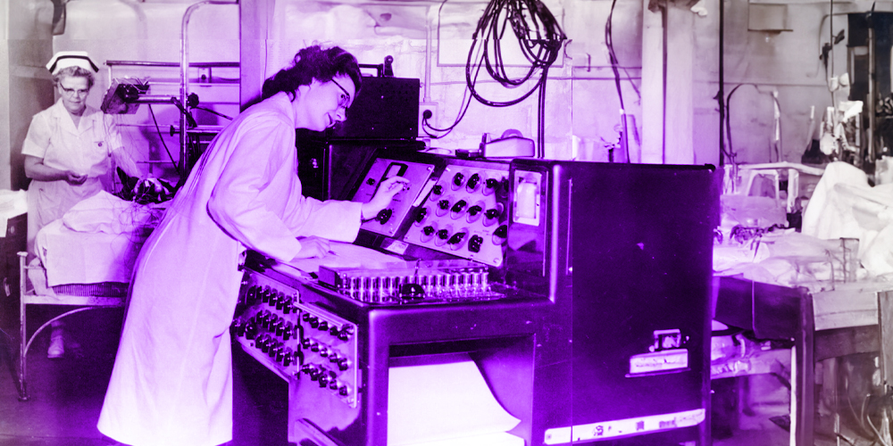
This technology, which includes techniques such as electroencephalography (EEG), electromyography (EMG), and evoked potentials, has evolved significantly since its inception in the early 20th century. The integration of these systems into clinical practice began gaining momentum in the 1970s and 1980s, improving surgical outcomes by enabling early detection of potential neural compromise.

The significance of neuromonitoring lies in its ability to enhance patient safety, reduce surgical risks, and optimize clinical decision-making by offering real-time neurophysiological feedback.

Furthermore, with advances in artificial intelligence (AI), cloud computing, and miniaturization, the future of neuromonitoring promises to enhance its precision, accessibility, and adaptability, further transforming its role in patient care.

This review explores the historical development, clinical applications, and future directions of neuromonitoring, highlighting its growing importance in modern medicine.

Introduction to Neuromonitoring

The origins of neuromonitoring date back to the early 20th century, with the invention of:

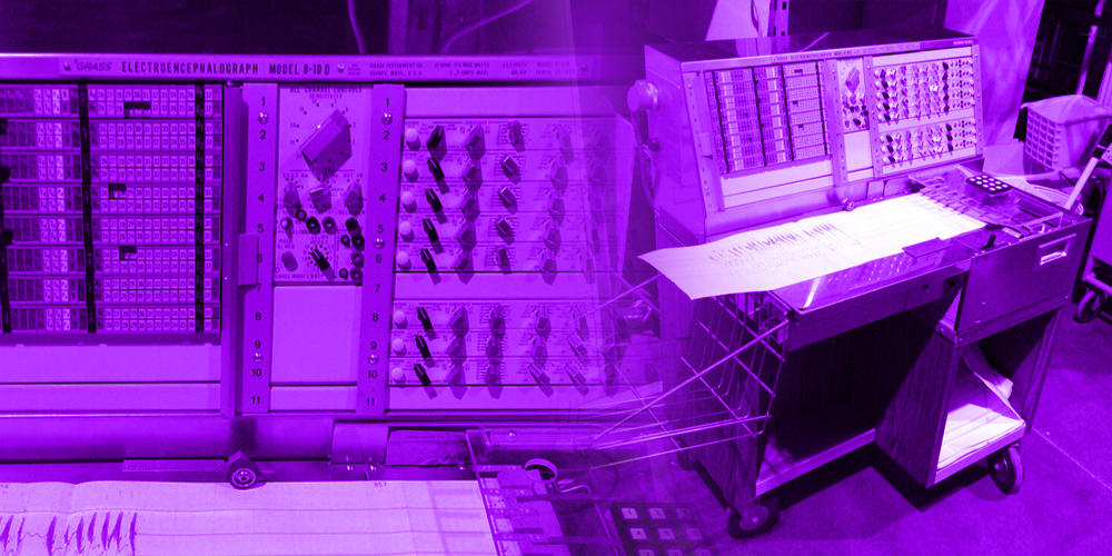
  • Electroencephalography (EEG) in the 1920s by Hans Berger, which provided a method to monitor electrical activity in the brain (1).
  • Electromyography (EMG) in the 1940s, allowing for the recording of muscle responses triggered by nerve stimulation (2).

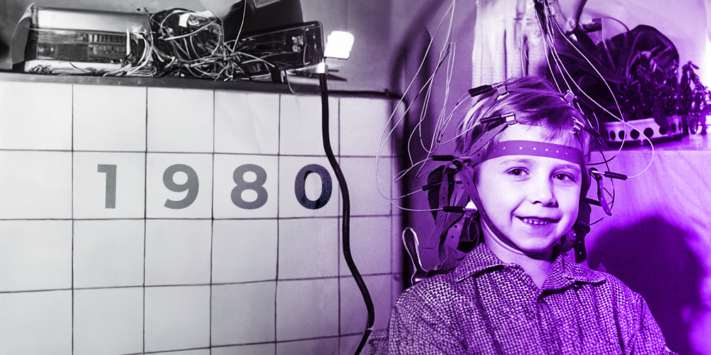
However, intraoperative neuromonitoring (IONM) gained widespread adoption in the 1970s and 1980s, following advancements in signal processing, neurophysiology, and instrumentation that made real-time monitoring feasible during complex surgeries (3).

Neuromonitoring is fundamentally rooted in the principles of neurophysiology, the branch of physiology that explores the functional properties of the nervous system at cellular, molecular, and systemic levels (4).

The nervous system operates through the generation and propagation of electrical impulses known as action potentials, which are initiated by ion exchange across neuronal membranes (4). These action potentials are essential for transmitting information between neurons and from neurons to muscles or sensory receptors via synaptic transmission (4).

In clinical neuromonitoring, these electrical signals are captured and interpreted using a range of techniques that exploit the bioelectrical nature of the nervous system.

Electroencephalography (EEG) records spontaneous electrical activity of the cerebral cortex and is especially valuable in detecting cortical dysfunction or seizure activity (5).

Electromyography (EMG) measures the electrical activity produced by skeletal muscles and is widely used to assess peripheral nerve integrity, particularly during spinal and cranial surgeries (6).

Evoked potentials (EPs), such as Somatosensory Evoked Potentials (SSEPs) and Motor Evoked Potentials (MEPs), are used to monitor the functional pathways of the central nervous system by applying a stimulus and recording the response (7).

SSEPs test the integrity of ascending sensory pathways, while MEPs evaluate descending motor pathways, especially useful in spinal and neurosurgical procedures (7).

Advanced neuromonitoring often integrates multimodal approaches, combining several neurophysiological techniques simultaneously to provide comprehensive feedback on the functional status of different components of the nervous system (8).

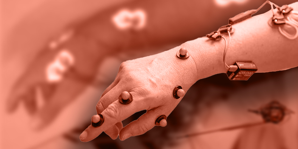
These techniques rely on both surface electrodes (placed on the scalp or skin) and needle electrodes (inserted intramuscularly or subdermally) for precise localization and quantification of neural activity.

Clinically, these signals serve as real-time indicators of neural integrity. A sudden loss or attenuation of an evoked potential, for example, may indicate ischemia, mechanical compression, or direct trauma to a neural structure (9).

This real-time feedback allows surgeons and anesthesiologists to adjust surgical technique, reposition instruments, or modify anesthesia protocols to mitigate the risk of permanent neurological injury (9). The integration of neurophysiological principles into intraoperative and critical care monitoring has therefore become essential for enhancing surgical safety, improving neurological outcomes, and reducing medico-legal risks (9).

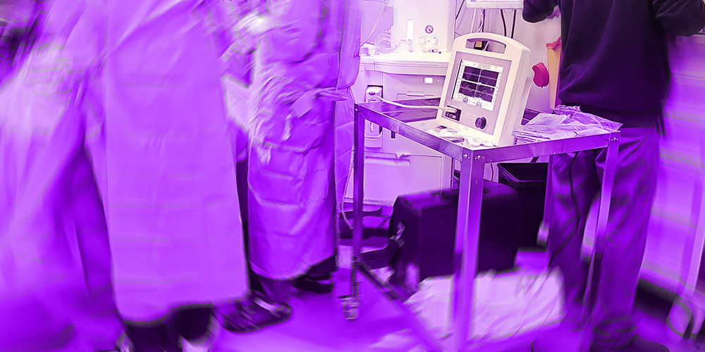
Neuromonitoring is a medical technology designed to assess and monitor the functional integrity of the nervous system in real-time during surgeries and critical care situations. It plays a vital role in neurosurgery, spinal surgery, ENT procedures, and critical care, enabling healthcare professionals to detect and prevent neurological damage before it becomes irreversible (9).

By continuously measuring electrical activity in the brain, spinal cord, and peripheral nerves, neuromonitoring enhances patient safety, surgical precision, and overall outcomes.

Definition of Neuromonitor

A neuromonitor, short for neurological monitor or neuromonitoring device, is a medical diagnostic tool designed to continuously assess and record electrical activity within the nervous system (10). It plays a critical role in detecting early signs of neurological impairment, especially during high-risk surgeries or in intensive care settings (11).

These devices are vital in ensuring the functional integrity of neural structures—such as the brain, spinal cord, and peripheral nerves—remains intact during procedures where these structures are vulnerable to injury (12).

Applications of Neuromonitors in Medicine

Neuromonitoring is utilized across a wide spectrum of clinical settings where real-time assessment of nervous system function is critical for patient safety. Its primary application is in the intraoperative environment, where it serves as a safeguard during surgeries that pose a risk to neural structures such as the brain, spinal cord, or peripheral nerves (2,8).

By continuously tracking neural signals, neuromonitoring helps surgeons detect early signs of nerve irritation or injury, enabling immediate corrective measures to prevent long-term deficits (3,8). Beyond the operating room, neuromonitoring is also employed in intensive care units (ICUs) to evaluate brain activity in patients with traumatic brain injury, stroke, or coma (11,13).

Additionally, it plays a key role in neurological diagnostics, such as epilepsy monitoring and assessing neuromuscular disorders (5,7). As such, neuromonitoring has become a cornerstone of modern neuroprotective strategies across various disciplines of medicine (9).

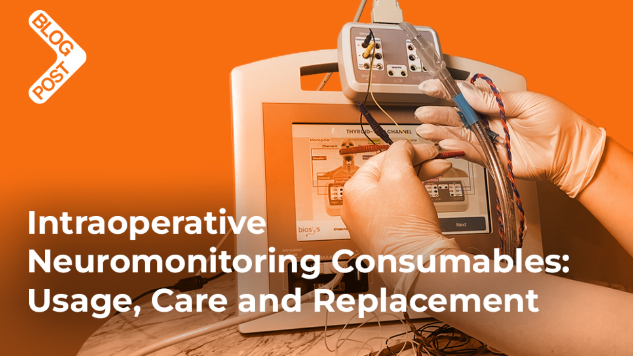
1. Intraoperative Neuromonitoring (IONM) in Surgery

Neuromonitors are extensively used during surgeries to monitor neural pathways and prevent neurological injuries. Their primary applications include:

  • Neurosurgery: Used for procedures such as brain tumor removal, epilepsy surgery, and aneurysm clipping, where preserving functional brain areas is essential (5,8).
  • Spinal Surgery: Ensures spinal cord integrity during procedures like scoliosis correction, spinal decompression, and spinal fusion surgeries using Somatosensory Evoked Potentials (SSEPs) and Motor Evoked Potentials (MEPs) (3,7,8).
  • Peripheral Nerve Surgery: Protects motor and sensory nerves during surgeries involving limbs or facial nerve reconstruction (8).
  • Vascular Surgery: Reduces the risk of stroke by monitoring brain activity and blood flow during carotid endarterectomy and aortic aneurysm repair (8).

Example: During spinal fusion surgery, SSEPs and MEPs are employed to continuously monitor the spinal cord’s functional integrity, minimizing the risk of postoperative neurological deficits (3,7).

2. Intensive Care Unit (ICU) & Neurocritical Monitoring

Neuromonitors are critical in the ICU for continuous assessment of brain function, particularly for patients with severe neurological conditions.

  • Traumatic Brain Injury (TBI): Monitoring intracranial pressure (ICP) and cerebral perfusion (11,13).
  • Stroke & Cerebral Ischemia: Evaluating blood flow and brain tissue oxygenation (13).
  • Coma & Brain Death Assessment: Using Continuous EEG (cEEG) to detect non-convulsive seizures or assess brain activity in unresponsive patients (11,13).
  • Brain Oxygenation Monitoring (PbtO₂, NIRS): Evaluating hypoxia and ischemia risks (13).

Example: In stroke patients, Transcranial Doppler (TCD) ultrasound is used to monitor cerebral blood flow and ensure adequate perfusion (13).

3. Epilepsy Monitoring & Diagnosis

Neuromonitors are vital tools in the diagnosis and management of epilepsy, particularly for patients with drug-resistant epilepsy.

  • Long-Term EEG Monitoring: Detects abnormal electrical activity and localizes seizure origins (5,7,14).
  • Video-EEG Monitoring: Simultaneously records physical symptoms and electrical activity during seizures (14).
  • Intracranial Depth Electrodes: Used for mapping seizure-prone areas in surgical candidates (14).

Example: Neuromonitoring helps identify the precise brain region responsible for seizure activity, guiding surgical removal of the affected area when medication is ineffective (14).

4. Anesthesia & Sedation Monitoring

Neuromonitoring is increasingly used to optimize anesthesia and sedation levels during surgery.

  • Bispectral Index (BIS) Monitoring: Measures depth of anesthesia to prevent under- or over-sedation (15).
  • Processed EEG (pEEG): Adjusts anesthesia dosage based on real-time brain activity (15).

Example: BIS monitoring ensures patients receive the appropriate level of anesthesia during high-risk neurosurgical procedures, enhancing recovery and minimizing side effects (15).

5. Neurological Research & Brain-Computer Interfaces (BCI)

Neuromonitors are essential for developing brain-machine interfaces (BMIs) and enhancing neuroprosthetic technologies.

  • Neuroprosthetics & Robotics: Allow paralyzed individuals to control external devices using brain signals (16).
  • AI-Driven Cognitive Monitoring: Facilitates early detection of dementia and other neurodegenerative conditions (16).
  • Neurofeedback therapy: Used in treating Attention-Deficit/Hyperactivity Disorder (ADHD), Post-Traumatic Stress Disorder (PTSD), and cognitive rehabilitation (16). Example: EEG-based BCIs allow individuals with severe disabilities to communicate and control assistive devices using brain signals alone (16).

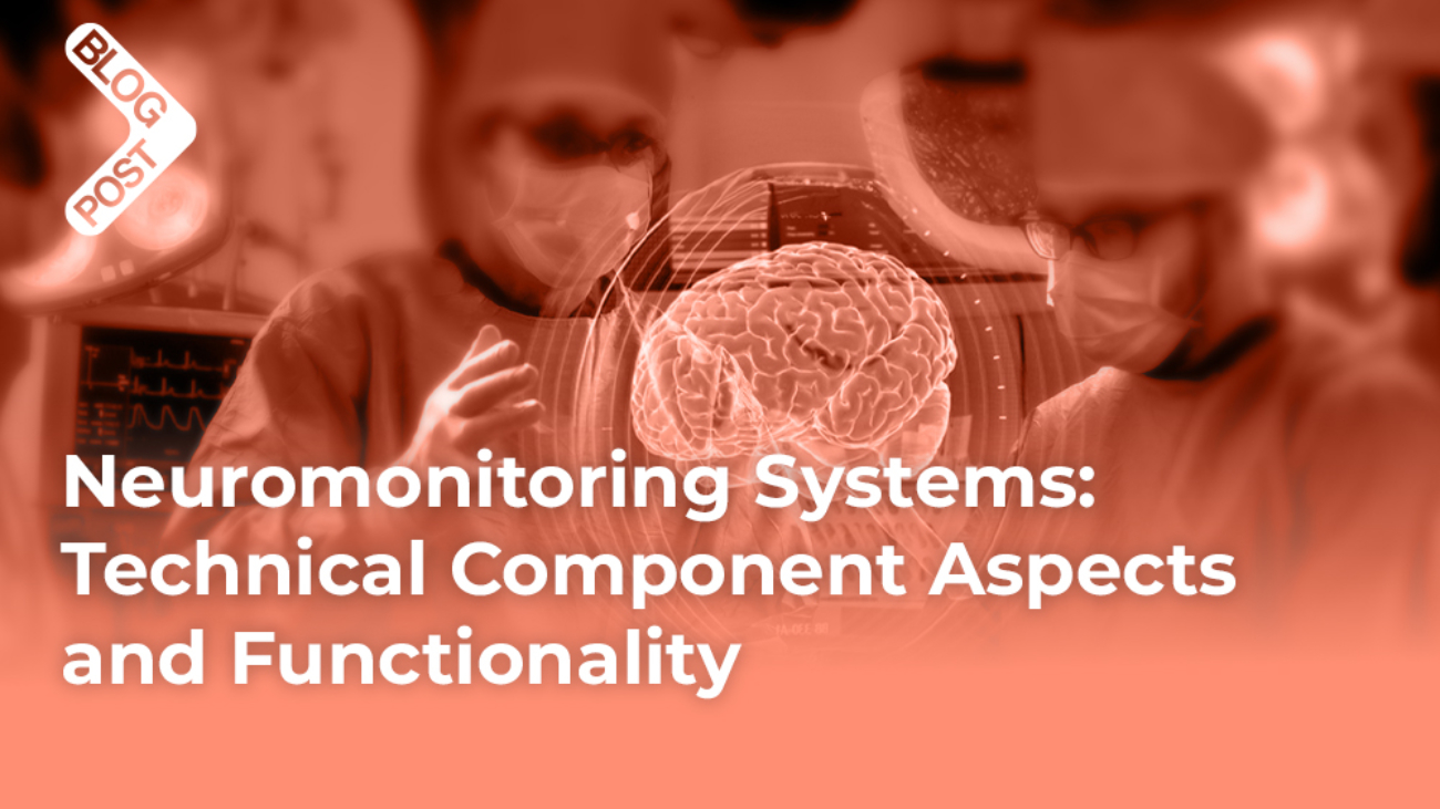
Technical Aspects of Neuromonitors

Neuromonitoring systems rely on a combination of sophisticated hardware and intelligent software to deliver real-time insights into neural function. From core sensors and signal processing techniques to intuitive interfaces and portable designs, each component plays a vital role in ensuring accurate, reliable, and clinically meaningful monitoring.

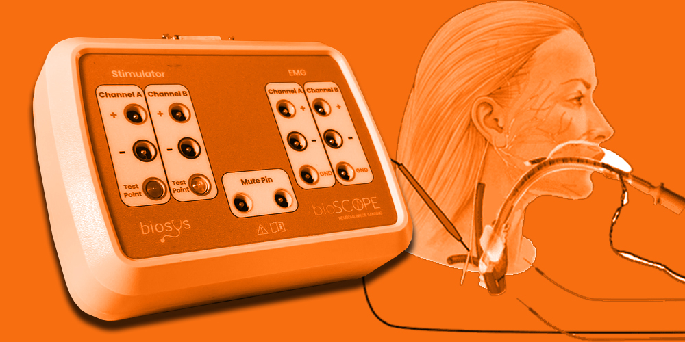
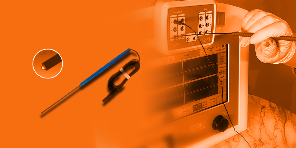
1. Core Sensors & Electrodes Used in Neuromonitoring

Neuromonitors employ various sensors to capture neural activity:

  • EEG Electrodes (Scalp & Depth Electrodes): Record brain electrical activity.
  • EMG Electrodes: Measure muscle activity to evaluate nerve integrity.
  • SSEP & MEP Electrodes: Stimulate and monitor sensory and motor pathways.
  • Near-Infrared Spectroscopy (NIRS): Non-invasively detects brain oxygenation.
  • Intracranial Pressure (ICP) Sensors: Monitor brain swelling in TBI patients.

2. Signal Processing & Interpretation

  • Amplification: Enhances weak neural signals for accurate interpretation.
  • Filtering: Removes artifacts caused by muscle movement, eye blinks, and external noise.
  • Machine Learning Algorithms: AI-based software identifies early-stage seizures, cerebral ischemia, and other anomalies.

Example: AI-enhanced EEG systems can predict seizures several hours before clinical symptoms appear, allowing for early interventions.

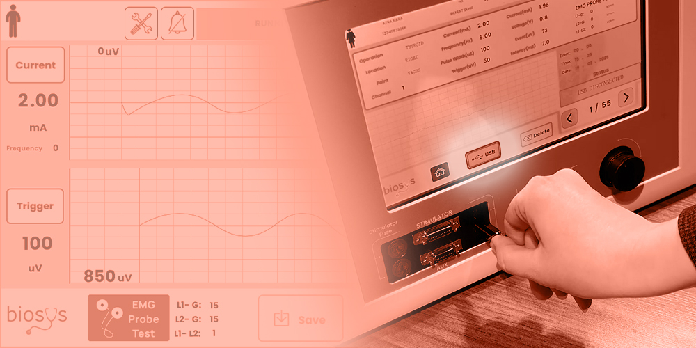
3. User Interface & Data Visualization

Modern neuromonitors offer intuitive interfaces that provide:

  • Real-time multi-channel waveforms.
  • Automated alerts for abnormal neural activity.
  • 3D Brain Mapping for surgical guidance.
  • Remote Monitoring via cloud-based AI.

Example: Portable neuromonitors like the Natus Brain Monitor provide continuous EEG tracking in ICUs, with instant alerts sent to clinicians’ mobile devices.

4. Wireless & Portable Neuromonitoring Devices

Technological advancements have led to portable and wireless systems that enhance patient accessibility:

  • Wearable EEG Headsets: Used for epilepsy monitoring and sleep disorder diagnosis.
  • Telemedicine-Enabled Diagnostics: Allows remote monitoring and care.
  • Wireless Neurostimulators: Assist patients with Parkinson’s disease and chronic pain.

Example: Bioscope by Biosys, initially a dual-channel neuromonitoring system for ENT, hand, and facial surgeries, is currently advancing to a 16-channel platform to expand its capabilities in both traditional and remote surgical environments.

Significance of Neuromonitoring

The nervous system, comprising the brain, spinal cord, and peripheral nerves, is highly vulnerable to injury during surgical interventions, trauma, or critical illness (4).

Even minimal damage to these structures can lead to devastating and often irreversible consequences such as motor deficits, sensory loss, chronic pain, paralysis, or cognitive dysfunction (4,9). This is particularly true during procedures involving delicate anatomical regions such as the spine, brainstem, cranial nerves, or peripheral nerve plexuses (8).

Intraoperative neuromonitoring (IONM) serves as a real-time surveillance system, enabling surgeons and anesthesiologists to continuously evaluate the functional integrity of neural pathways during surgical procedures (3,8).  

By detecting subtle changes in neural activity, IONM provides early warning signs of potential nerve compromise before structural damage becomes permanent (8). This allows the surgical team to immediately adjust their technique—such as repositioning surgical tools, reducing traction, or altering the depth of anesthesia—to prevent injury (3,8).

Furthermore, neuromonitoring contributes to improved functional and neurological outcomes. Numerous clinical studies have shown that the use of IONM in spine and brain surgeries significantly reduces the incidence of postoperative neurological deficits (9,17).  

For instance, in scoliosis correction or tumor resection surgeries, monitoring somatosensory evoked potentials (SSEPs) and motor evoked potentials (MEPs) helps ensure spinal cord integrity throughout the operation (7,9,17).

In critical care settings, such as the intensive care unit (ICU), neuromonitoring technologies like EEG and bispectral index (BIS) allow clinicians to assess brain function in patients with coma, traumatic brain injury, or under sedation (11,13,15).  

This enables timely interventions that may mitigate secondary brain injury, optimize ventilator management, or evaluate seizure activity (11,13,15).

In summary, the importance of neuromonitoring lies in its ability to:

  • Enhance surgical safety by reducing the risk of intraoperative nerve damage (8,9).
  • Support clinical decision-making with real-time neurophysiological feedback (8,9).
  • Improve prognosis and long-term outcomes by preserving neurological function (9,17).
  • Reduce healthcare burden by preventing complications that may lead to prolonged hospitalization, rehabilitation, or lifelong disability (17).
  • Improves Surgical Precision: Guides surgeons to avoid critical nerve pathways (8,9).
  • Reduces Post-Surgical Complications: Minimizes risks of paralysis and cognitive impairment (9,17).
  • Supports Predictive Medicine: AI-enhanced systems enable early diagnosis of neurodegenerative diseases (16).
  • Enhances Patient Recovery: Facilitates faster rehabilitation (9,17).

As surgical procedures become more complex and patient populations more vulnerable, the role of neuromonitoring becomes increasingly essential in delivering high-quality, patient-centered care (9,17).

Future of Neuromonitoring

Neuromonitoring is undergoing a transformative evolution driven by technological advancements, with the future promising smarter, more adaptive, and more accessible monitoring solutions (16,18). As surgical procedures grow increasingly complex and precision becomes paramount, the demand for advanced neuromonitoring capabilities has never been greater (9,18).

Future directions in this field are largely shaped by the integration of artificial intelligence (AI), machine learning, cloud-based platforms, and real-time remote monitoring, all of which aim to enhance safety, efficiency, and patient outcomes (16,18,19).

1. Artificial Intelligence and Predictive Analytics

AI is beginning to revolutionize how neuromonitoring data is interpreted. By applying machine learning algorithms to large datasets of neurophysiological signals, systems can identify subtle patterns and deviations that may precede neurological injury (16,18). This allows for predictive diagnostics, enabling clinicians to act before critical thresholds are crossed. For example, AI-enhanced systems can detect early signs of ischemia or nerve traction injuries based on changes in signal amplitude or latency long before these would be apparent to the human eye (16,18,19).

2. Cloud-Based Data Storage and Analysis

The integration of cloud computing facilitates secure, centralized storage and analysis of neuromonitoring data (20). This approach offers several benefits, including:

  • Long-term tracking of patient neurological trends (20).
  • Facilitating multi-center collaborations and comparative studies (20).
  • Providing remote access to data for expert consultations, even during surgery (20).
  • Enabling real-time audits and quality assurance for improved accountability and outcomes (20).

3. Remote and Telemonitoring Capabilities

With the rise of telemedicine, neuromonitoring systems are beginning to support remote supervision (21). This allows expert neurophysiologists to oversee and interpret intraoperative neuromonitoring from different locations, enhancing the availability of specialized care in rural or underserved hospitals (21). This development also supports the scalability of neuromonitoring services, ensuring that high-risk procedures can be safely conducted even in lower-resourced settings (21).

4. Miniaturization and Wearable Neuromonitors

The development of portable and wearable neuromonitoring devices is another key innovation on the horizon (22). These compact systems can continuously monitor neural activity outside the operating room — such as in ICU patients, during sleep studies, or in home rehabilitation settings — offering clinicians new ways to manage and follow neurological function over time (22).

5. Enhanced User Interfaces and Automation

Modern neuromonitoring systems are being designed with more intuitive user interfaces, automatic signal calibration, and smart artifact reduction features (19,22). These improvements reduce the cognitive and technical load on operating room staff and improve the accuracy and reliability of the recorded data (19,22).

6. Integration with Surgical Robotics and Navigation

As robot-assisted surgeries become more common, future neuromonitoring systems are being designed to integrate seamlessly with robotic platforms and navigation systems (23). This allows real-time neurophysiological feedback to directly guide robotic movement, reducing human error and increasing surgical precision (23).

The future of neuromonitoring is geared toward making surgeries safer, more efficient, and more patient-specific (18,19). With ongoing advances in AI, connectivity, hardware, and interface design, neuromonitoring is poised to become an even more integral part of precision medicine and surgical care (16,18,19).

These innovations will not only enhance intraoperative decision-making but also expand the reach of neuromonitoring into diagnostics, rehabilitation, and long-term neurological care (16,19,22).

References

  1. Berger, H. (1929). Über das Elektrenkephalogramm des Menschen. Archiv für Psychiatrie und Nervenkrankheiten, 87(1), 527–570. https://doi.org/10.1007/BF01797193
  2. Preston, D. C., & Shapiro, B. E. (2013). Electromyography and neuromuscular disorders: Clinical-electrophysiologic correlations (3rd ed.). Elsevier.
  3. MacDonald, D. B. (2006). Intraoperative motor evoked potential monitoring: Overview and update. Journal of Clinical Monitoring and Computing, 20(5), 347–377. https://doi.org/10.1007/s10877-006-9040-6
  4. Purves, D., Augustine, G. J., & Fitzpatrick, D. (2018). Neuroscience (6th ed.). Oxford University Press.
  5. Niedermeyer, E., & da Silva, F. L. (2004). Electroencephalography: Basic principles, clinical applications, and related fields (5th ed.). Lippincott Williams & Wilkins.
  6. Dumitru, D., Amato, A. A., & Zwarts, M. J. (2002). Electrodiagnostic medicine (2nd ed.). Hanley & Belfus.
  7. Nuwer, M. R. (1998). Intraoperative monitoring of neural function. In Handbook of Clinical Neurophysiology (Vol. 3, pp. 841–871). Elsevier.
  8. Sala, F., & Deletis, V. (2007). Intraoperative neurophysiology in neurosurgery: A short overview. Clinical Neurophysiology, 118(3), 525–528. https://doi.org/10.1016/j.clinph.2006.10.021
  9. Skinner, S. A., Transfeldt, E. E., & Grafton, S. T. (2008). Intraoperative neurophysiologic monitoring: A review of techniques used to monitor the nervous system during spine surgery. Neurosurgical Focus, 25(3), E6. https://doi.org/10.3171/FOC/2008/25/9/E6
  10. MacDonald, D. B., Skinner, S., Shils, J., & Yingling, C. (2013). Intraoperative motor evoked potential monitoring – A position statement by the American Society of Neurophysiological Monitoring. Clinical Neurophysiology, 124(12), 2291–2316. https://doi.org/10.1016/j.clinph.2013.07.025
  11. Oddo, M., Bösel, J., Helbok, R., et al. (2018). Monitoring the injured brain: ICP and advanced neuromonitoring. Intensive Care Medicine, 44(12), 1888–1890. https://doi.org/10.1007/s00134-018-5448-2
  12. Nuwer, M. R., Dawson, E. G., Carlson, L. G., & Kanim, L. E. (1995). Somatosensory evoked potential spinal cord monitoring reduces neurologic deficits after scoliosis surgery: Results of a large multicenter survey. Electroencephalography and Clinical Neurophysiology, 96(1), 6–11. https://doi.org/10.1016/0013-4694(95)00035-6
  13. Smith, S. J. M. (2005). EEG in the diagnosis, classification, and management of patients with epilepsy. Journal of Neurology, Neurosurgery & Psychiatry, 76(suppl 2), ii2–ii7. https://doi.org/10.1136/jnnp.2005.069245
  14. Rampil, I. J. (1998). A primer for EEG signal processing in anesthesia. Anesthesiology, 89(4), 980–1002. https://doi.org/10.1097/00000542-199810000-00023
  15. Lebedev, M. A., & Nicolelis, M. A. L. (2006). Brain–machine interfaces: past, present and future. Trends in Neurosciences, 29(9), 536–546. https://doi.org/10.1016/j.tins.2006.07.004
  16. Chuang, C. F., Chen, Y. J., Chang, Y. J., et al. (2021). Artificial intelligence applications in intraoperative neuromonitoring: A review. Frontiers in Medicine, 8, 655105. https://doi.org/10.3389/fmed.2021.655105
  17. Fehlings, M. G., Brodke, D. S., Norvell, D. C., & Dettori, J. R. (2010). The evidence for intraoperative neurophysiological monitoring in spine surgery: Does it make a difference? Spine, 35(9 Suppl), S37–S46. https://doi.org/10.1097/BRS.0b013e3181d82c74
  18. Varatharajan, R., & Navaneethakrishnan, S. (2020). Artificial intelligence based neural monitoring and robotics in surgery. Surgical Innovation, 27(6), 663–673. https://doi.org/10.1177/1553350620943535
  19. El-Osta, A., Elghazaly, A., & Said, M. (2022). Cloud computing in healthcare: Review and research challenges. Healthcare Technology Letters, 9(2), 45–53. https://doi.org/10.1049/htl2.12016
  20. Garcia, R. M., & Sherman, E. M. (2020). Tele-neuromonitoring: Intraoperative neurophysiological monitoring from a distance. Clinical Neurophysiology, 131(1), 305–312. https://doi.org/10.1016/j.clinph.2019.10.022
  21. Khan, M. J., Hong, M. J., & Hong, K. S. (2018). Decoding of four movement directions using hybrid NIRS-EEG brain-computer interface. Frontiers in Human Neuroscience, 12, 244. https://doi.org/10.3389/fnhum.2018.00244
  22. Baek, H., Chung, J., Kim, Y. J., & Park, Y. S. (2021). Real-time neurophysiological monitoring during robot-assisted neurosurgery. Frontiers in Surgery, 8, 658776. https://doi.org/10.3389/fsurg.2021.658776
Blog_Cover_

IONM Consumables: Usage, Care and Replacement

The Bioscope by Biosys system requires various consumables to function effectively during ENT, hand, and facial surgeries. Here’s how to properly use, maintain, and replace these consumables, followed by a comparison with brand A and brand B.

How to Properly Use, Maintain, and Replace Bioscope Consumables

1. Needle Electrodes (EMG Monitoring)

  • Use:
    • Insert needle electrodes into the target muscle or nerve area to record EMG signals.
    • Ensure proper placement to avoid inaccurate readings or tissue damage.
  • Maintenance:
    • None; these are single-use only.
  • Replacement:
    • Discard after each surgery following biohazard disposal protocols.
    • Cost per electrode: $5 – $20.

2. Adhesive Surface Electrodes (EMG, SSEP Monitoring)

  • Use:
    • Apply a thin layer of conductive gel or paste to improve signal conductivity.
    • Place electrodes over the skin where nerve signals are to be detected (e.g., vocal cords or facial nerves).
    • Secure electrodes properly to prevent shifting during surgery.
  • Maintenance:
    • None; single-use only.
  • Replacement:
    • Discard after each surgery.
    • Cost per set: $10 – $50.

3. Electrode Pads (Scalp or Skin Contact)

  • Use:
    • Attach to the skin using adhesive backing.
    • Ensure proper placement to minimize signal loss.
  • Maintenance:
    • None; disposable and single-use.
  • Replacement:
    • Replace after each use.
    • Cost per set: $10 – $30.

4. Conductive Gel and Paste

  • Use:
    • Apply directly to the skin before attaching electrodes.
    • Spread evenly to ensure good electrical conductivity.
  • Maintenance:
    • Store in a cool, dry place with the cap tightly sealed.
  • Replacement:
    • Replace after each patient use.
    • Cost per tube: $5 – $15.

5. Disposable Stimulation Probes (If Used)

  • Use:
    • Apply sterile stimulation probes to nerve locations during stimulation tests.
    • Ensure precise placement for effective nerve response monitoring.
  • Maintenance:
    • None; single-use only.
  • Replacement:
    • Discard after each procedure.
    • Cost per probe: $50 – $200.

6. Connecting Cables (Limited-Use)

  • Use:
    • Connect electrodes to the amplifier and processing unit.
    • Ensure proper connection points to prevent signal disruption.
  • Maintenance:
    • Clean cables with alcohol wipes after each use.
    • Inspect for wear, fraying, or loss of insulation.
  • Replacement:
    • Replace when damaged or signal integrity is compromised.
    • Cost per cable: $20 – $100.

7. Sterile Drapes & Covers (Protective Use)

  • Use:
    • Cover non-sterile components to maintain a sterile field.
    • Protect electrodes and cables from contamination.
  • Maintenance:
    • None; single-use only.
  • Replacement:
    • Discard after each procedure.
    • Cost per drape or cover: $5 – $15.

Comparison with Brand A & Brand B IONM Systems

AspectBioscope by BiosysBrand ABrand B
Needle ElectrodesSingle-use, disposable.Single-use, disposable.Single-use, disposable.
Adhesive ElectrodesSingle-use, disposable.Single-use, disposable.Single-use, disposable.
Electrode PadsSingle-use, disposable.Reusable types available for EEG.Reusable and single-use options.
Conductive Gel/PasteSingle-use per patient.Single-use per patient.Single-use per patient.
Stimulation ProbesSingle-use, limited-use.Reusable with sterilization.Reusable and disposable options.
Connecting CablesReusable, limited lifespan.Reusable, more durable.Reusable, high-quality shielding.
Sterile Drapes & CoversSingle-use.Single-use.Single-use.
Cost Range (Per Use)$100 – $300 per surgery.$200 – $500 per surgery.$250 – $600 per surgery.
Sterilization NeedsLimited to connecting cables and stimulation probes.Extensive for probes and cables.Extensive for probes, cables, and electrodes.
Maintenance FrequencyLow to moderate.High (more parts requiring sterilization).High (multi-modality systems).
System FocusENT & Peripheral Nerve Monitoring.Cranial, Spinal, and ENT Monitoring.Comprehensive, Multi-Modality Monitoring.

The Bioscope by Biosys, designed for simplicity and cost-effectiveness, is ideal for ENT, hand, and facial surgeries due to its low maintenance and minimal consumable requirements. It provides essential monitoring, especially for laryngeal nerve function, making it a valuable tool in delicate nerve surgeries. Its ease of use and affordability make it a go-to option for institutions with limited resources.

Brand A offers multi-modality support, including EEG, EMG, MEP, and SSEP monitoring capabilities, making it versatile in a wide range of surgical procedures, from cranial to spinal surgeries. While it uses similar consumables to other systems, its complex nature demands more thorough sterilization and frequent maintenance. The system’s multi-modality capabilities make it invaluable in high-risk surgeries but also require more careful handling to ensure reliability and patient safety.

Brand B, a comprehensive and high-precision system, is the most resource-intensive of the three. It supports a wide variety of surgeries, including spinal and brain procedures, and requires frequent replacement of high-quality electrodes and probes. Its advanced features make it suitable for complex surgeries, but the broad application scope comes with the tradeoff of higher operational costs and greater maintenance needs.

Standard Operating Procedure (SOP) for Using, Maintaining, and Replacing Consumables in the Bioscope System

This SOP will help you ensure effective use, maintenance, and replacement of consumables for the Bioscope by Biosys neuromonitoring system. It also includes cost optimization strategies and efficiency improvements.

Section 1: Preparation & Setup

1.1 Preparation Before Surgery

  • Check Inventory:
    • Ensure sufficient supply of all required consumables:
      • Needle Electrodes (EMG)
      • Adhesive Surface Electrodes (EMG, SSEP)
      • Electrode Pads
      • Conductive Gel / Paste
      • Disposable Stimulation Probes (if used)
      • Connecting Cables (reusable)
      • Sterile Drapes & Covers
  • Visual Inspection:
    • Examine reusable components (cables, stimulation probes) for signs of damage, fraying, or corrosion.
    • Replace damaged items immediately.
  • Sterility Check:
    • Confirm that all disposable items are sealed and sterile before use.
  • Equipment Calibration:
    • Test and calibrate the signal amplifier and data acquisition unit.
    • Ensure all connections are secure and functional.

Section 2: Intraoperative Use

2.1 Setting Up Consumables

  • Needle Electrodes (EMG):
    • Insert into the muscle groups associated with the nerves being monitored.
    • Secure using adhesive pads or clips as needed.
  • Adhesive Surface Electrodes (EMG, SSEP):
    • Apply a thin layer of conductive gel or paste to the skin before placement.
    • Attach electrodes firmly and ensure proper contact.
  • Electrode Pads:
    • Apply directly to the skin where signal acquisition is needed (e.g., scalp or limbs).
  • Stimulation Probes (If Used):
    • Attach to the nerve sites for direct stimulation.
    • Monitor response via the Bioscope interface for real-time feedback.

Section 3: Post-Surgery Cleanup & Maintenance

3.1 Consumable Replacement

  • Single-Use Components:
    • Discard all used needle electrodes, adhesive electrodes, electrode pads, stimulation probes, and drapes/covers in appropriate biohazard disposal containers.
  • Limited-Use Components (Connecting Cables):
    • Clean cables with alcohol wipes or disinfectant solutions.
    • Inspect cables for physical damage before next use.

3.2 Sterilization of Reusable Components

  • Connecting Cables & Stimulation Probes:
    • Clean with 70% isopropyl alcohol wipes or appropriate disinfectants.
    • For stimulation probes:
      • If reusable, sterilize using an autoclave (if heat-tolerant) or chemical disinfectants (e.g., glutaraldehyde).
    • Dry thoroughly before storing.

3.3 Data Backup & Software Maintenance

  • Save patient data to secure storage systems as per medical standards.
  • Check for software updates to maintain optimal performance.
  • Run diagnostic tests to ensure proper calibration and system integrity.

Section 4: Cost Optimization & Efficiency Improvements

Strategies to Reduce Costs and Improve Efficiency

  1. Bulk Purchasing:
    • Purchase needle electrodes, adhesive electrodes, and electrode pads in bulk to reduce per-unit costs.
  2. Using Reusable Components Where Safe:
    • Invest in high-quality, reusable connecting cables and stimulation probes.
    • Follow strict sterilization protocols to maximize lifespan.
  3. Maintenance Schedule:
    • Implement a scheduled maintenance plan for reusable components to ensure reliability and reduce replacement frequency.
  4. Regular Supplier Evaluation:
    • Compare suppliers for competitive pricing and quality.
    • Negotiate contracts with manufacturers for long-term supply at reduced rates.
  5. Staff Training:
    • Provide training on proper handling, cleaning, and disposal procedures to prevent damage and contamination.

Section 5: Comparison with Other Systems

AspectBioscope by BiosysBrand ABrand B
Single-Use Parts CostLower (Approx. $100 – $300 per surgery)Moderate (Approx. $200 – $500 per surgery)High (Approx. $250 – $600 per surgery)
Sterilization NeedsLimited to cables and stimulation probesExtensive; requires frequent sterilization of probes and cables.Frequent sterilization needed for all components.
Reusable ComponentsCables, Stimulation Probes (if used)Cables, Probes, Certain ElectrodesCables, Electrodes, Stimulation Devices
Maintenance FrequencyModerate (Few components to sterilize)High (Multiple components)High (Multi-modality systems)
Cost Optimization MethodsBulk purchasing and reusing cables where possible.Long-term contracts with suppliers.Careful maintenance and cleaning of reusable parts.
Component LifespanModerate to High (Reusable components last longer with proper care).Moderate (High maintenance cost).High (Excellent durability but complex handling).

Use of Neuromonitoring in ENT (Ear, Nose, Throat) Operations

Neuromonitoring is increasingly used in ENT surgeries to preserve nerve integrity and prevent nerve damage, especially when critical nerves are at risk of injury. Its use in ENT procedures is particularly important because even minor damage to nerves can cause significant functional impairments, such as voice loss, swallowing difficulties, or facial paralysis.

Common ENT Surgeries Where Neuromonitoring Is Used

1. Thyroid and Parathyroid Surgery (Most Common Use)

  • Nerve at Risk: Recurrent Laryngeal Nerve (RLN) and Superior Laryngeal Nerve (SLN).
  • Why Neuromonitoring is Important:
    • The RLN controls the muscles of the vocal cords; injury can cause hoarseness, loss of voice, or breathing difficulties.
    • The SLN affects voice pitch; injury can result in difficulty modulating voice.
  • How Neuromonitoring is Used:
    • Electrodes are placed on the vocal cord muscles (via endotracheal tube electrodes) or on the skin near the laryngeal muscles.
    • Electrical stimulation is applied, and the EMG response is monitored in real-time.
    • Visual and audio alerts indicate proximity to or damage of the nerve.

2. Parotid Gland Surgery (Facial Nerve Monitoring)

  • Nerve at Risk: Facial Nerve (CN VII).
  • Why Neuromonitoring is Important:
    • The Facial Nerve controls muscle movements of the face.
    • Injury can cause partial or complete facial paralysis.
  • How Neuromonitoring is Used:
    • Surface or needle electrodes are placed in facial muscles innervated by the facial nerve.
    • Continuous EMG monitoring detects nerve activity during surgery.
    • Alerts warn the surgeon when nerve manipulation is detected.

3. Skull Base Surgery (Cranial Nerve Monitoring)

  • Nerves at Risk: Facial Nerve (CN VII), Vagus Nerve (CN X), Glossopharyngeal Nerve (CN IX).
  • Why Neuromonitoring is Important:
    • The nerves involved are essential for swallowing, speech, facial movements, and other critical functions.
    • Damage can cause permanent deficits if not detected early.
  • How Neuromonitoring is Used:
    • Electrode placement around nerves at risk.
    • Monitoring of muscle activity during surgical manipulation.
    • Audio-visual feedback provided in real-time.

4. Cochlear Implant Surgery

  • Nerve at Risk: Facial Nerve (CN VII).
  • Why Neuromonitoring is Important:
    • The facial nerve is very close to the surgical area when inserting cochlear implants.
    • Prevents accidental damage during electrode array insertion.
  • How Neuromonitoring is Used:
    • Continuous monitoring of facial muscle activity to avoid nerve damage.
    • EMG electrodes are placed over relevant muscles.

5. Ear Surgeries (e.g., Mastoidectomy)

  • Nerve at Risk: Facial Nerve (CN VII).
  • Why Neuromonitoring is Important:
    • Injury to the facial nerve can result in facial droop or paralysis.
    • Necessary during procedures involving the mastoid bone or middle ear.
  • How Neuromonitoring is Used:
    • Real-time monitoring of nerve function using EMG electrodes.
    • Electrical stimulation can be applied to detect nerve presence.

How Neuromonitoring Works in ENT Surgeries (Bioscope Example)

  1. Placement of Electrodes:
    • Needle or surface electrodes are placed on or near the muscles controlled by the nerves at risk (e.g., laryngeal muscles for RLN monitoring).
  2. Nerve Stimulation:
    • Low-level electrical stimulation is applied near the surgical area.
    • The nerve response is recorded by the electrodes.
  3. Signal Interpretation:
    • The recorded signals are processed by the Bioscope system.
    • Waveforms are displayed on the touchscreen and provide visual/audio alerts.
  4. Safety Alerts:
    • When nerve damage or proximity is detected, the system provides immediate feedback to the surgical team.

Comparison with Other Neuromonitoring Systems

SystemBioscope by BiosysBrand ABrand B
Primary ApplicationENT (Thyroid, Parathyroid, Facial Surgeries)ENT, Cranial, Spinal SurgeriesComprehensive (ENT, Spinal, Brain Surgeries)
Nerves MonitoredRLN, SLN, Facial NerveCranial & Spinal NervesPeripheral, Cranial & Spinal Nerves
Alert SystemsVisual & Audio AlertsVisual & Audio AlertsVisual & Audio Alerts
Display Type10.1-inch TouchscreenStandard LED/TouchscreenHigh-Resolution Display
PortabilityYes (Battery-Powered)NoYes (Limited)
Cost EfficiencyMore Cost-Effective for ENT ApplicationsModerate to HighHigh

Benefits of Using Neuromonitoring in ENT (Ear, Nose, Throat) Surgeries

Neuromonitoring has become a critical tool in ENT surgeries, where preserving nerve function is essential to maintaining speech, facial movements, breathing, swallowing, and hearing. The implementation of neuromonitoring devices, such as the Bioscope by Biosys, provides significant benefits across various ENT procedures.

1. Enhanced Nerve Safety and Preservation

  • ENT surgeries often involve high-risk nerves like the Recurrent Laryngeal Nerve (RLN), Superior Laryngeal Nerve (SLN), and Facial Nerve (CN VII).
  • Real-time monitoring allows surgeons to detect and avoid damage to these nerves during procedures like:
    • Thyroidectomy and Parathyroidectomy (RLN and SLN monitoring).
    • Parotid Gland Surgery (Facial Nerve monitoring).
    • Cochlear Implantation and Mastoidectomy (Facial Nerve monitoring).
  • Prevents permanent functional loss, including:
    • Hoarseness or voice loss (RLN injury).
    • Pitch modulation difficulties (SLN injury).
    • Facial paralysis or drooping (Facial Nerve injury).

2. Increased Surgical Precision

  • Visual and Audio Feedback:
    • Systems like Bioscope provide real-time visual displays and audio alerts when nerves are at risk.
    • Surgeons can make precise adjustments instantly based on the feedback received.
  • Better Identification of Nerve Structures:
    • Improves detection of nerves that are difficult to visualize due to scar tissue, tumors, or anatomical variations.
    • Enhances precision in tumor resections, parotidectomies, and complex ear surgeries.

3. Reduced Postoperative Complications

  • Significantly reduces the risk of:
    • Vocal cord paralysis during thyroid and parathyroid surgeries.
    • Facial muscle dysfunction following parotid and skull base surgeries.
    • Swallowing difficulties and airway compromise from nerve damage.
  • Faster recovery times due to reduced nerve trauma and improved surgical technique.

4. Improved Surgical Outcomes

  • Enhanced nerve preservation leads to better functional recovery and patient satisfaction.
  • Reduced rates of reoperation due to nerve damage.
  • Minimizes the risk of long-term disabilities associated with nerve injuries.

5. Early Detection of Nerve Stress or Injury

  • Continuous monitoring during surgery helps identify early signs of nerve stress or dysfunction.
  • Systems like Bioscope alert the surgeon before permanent damage occurs, allowing for immediate correction.
  • Detection of near-miss injuries prevents complications that may only become evident postoperatively.

6. Cost-Efficiency in Preventing Complications

  • By preventing nerve damage, neuromonitoring can save significant healthcare costs associated with:
    • Prolonged hospital stays.
    • Additional surgeries required to repair nerve damage.
    • Long-term rehabilitation or therapy costs.
  • Bioscope’s focus on ENT procedures provides a cost-effective solution specifically tailored for laryngeal and facial nerve monitoring.

7. Enhanced Training and Surgical Education

  • Neuromonitoring systems provide visual aids and feedback that can be used for educational purposes.
  • Surgeons in training can learn precise nerve identification and protection techniques with real-time guidance.

8. Better Patient Communication and Satisfaction

  • The use of neuromonitoring can be explained to patients as a safety measure that enhances surgical precision and reduces risks.
  • Improved patient confidence in the procedure knowing that measures are in place to protect critical nerves.

9. Minimally Invasive Surgery Compatibility

  • Neuromonitoring systems like Bioscope can be integrated with minimally invasive surgical techniques, offering benefits such as:
    • Smaller incisions.
    • Reduced tissue trauma.
    • Faster healing and recovery.

10. Integration with Other Surgical Technologies

  • Neuromonitoring systems can work in tandem with:
    • Surgical microscopes.
    • Endoscopic instruments.
    • Robotic-assisted surgical tools.
  • Enhances the accuracy of delicate procedures where visualization alone is not sufficient.

Comparison of Benefits: Bioscope vs. Other Systems

BenefitBioscope by BiosysBrand ABrand B
Real-Time FeedbackVisual & Audio AlertsVisual & Audio AlertsVisual & Audio Alerts
Cost EfficiencyHigh (ENT-Focused)Moderate (Multi-Purpose)Low (Comprehensive Monitoring)
Ease of UseHigh (User-Friendly UI)Moderate (More Complex Setup)Moderate (Advanced System)
PortabilityHigh (Battery-Powered)Low (Stationary Systems)Moderate (Limited Portability)
Best ApplicationsENT Surgeries (RLN, SLN, Facial Nerve)ENT, Spinal SurgeriesComprehensive Nerve Monitoring
Customization OptionsLimited (Dual-Channel)High (Multi-Modality)High (Multi-Modality)
Blog_Cover_Template_07.05.2025

Neuromonitoring Systems: Technical Component Aspects and Functionality

A neuromonitoring system (neuromonitor) is a sophisticated medical device designed to assess the integrity and functionality of the nervous system in real-time.
It consists of several interdependent technical components that work in harmony to detect, amplify, process, and interpret neural signals during surgical procedures, critical care, and diagnostic evaluations.

The core elements of a neuromonitoring system can be categorized into hardware, software, and user interfaces, each contributing to the accuracy, reliability, and clinical utility of the system.
Understanding these components is crucial to appreciating how neuromonitoring enhances patient safety and surgical precision.

Main Parts of a Neuromonitoring System

  1. Electrodes & Sensors (Signal Detection)
    • EEG Electrodes:
      • Detect brain electrical activity, usually placed on the scalp.
      • Types: Scalp electrodes, Depth electrodes (for invasive monitoring).
    • EMG Electrodes (Electromyography):
      • Measure muscle activity to evaluate nerve integrity.
      • Typically used for monitoring cranial nerves and peripheral nerves.
    • SSEP Electrodes (Somatosensory Evoked Potentials):
      • Assess sensory pathway integrity by recording neural responses to sensory stimulation (e.g., electrical, tactile).
      • Often used in spinal surgeries.
    • MEP Electrodes (Motor Evoked Potentials):
      • Monitor motor pathway integrity by detecting muscle responses following nerve stimulation.
      • Essential in procedures where motor nerve function is at risk.
    • Near-Infrared Spectroscopy (NIRS):
      • Measures brain oxygenation levels non-invasively.
      • Commonly used in ICU settings and brain surgeries.
    • Intracranial Pressure (ICP) Sensors:
      • Monitor pressure within the skull, especially important in trauma or brain injury management.
  1. Stimulators (Signal Generation)
    • Electrical Stimulators:
      • Provide precise electrical pulses to stimulate nerves for evoked potential monitoring (MEPs, SSEPs).
      • Can be surface-based or needle-based depending on application.
    • Transcranial Magnetic Stimulators (TMS):
      • Non-invasive method of stimulating cortical motor areas.
      • Useful in mapping functional brain areas during surgery.
  1. Signal Amplifiers (Signal Processing)
    • High-Gain Amplifiers:
      • Boost weak neural signals to measurable levels without distortion.
      • Essential for processing EEG, EMG, SSEP, and MEP signals.
    • Noise Filters:
      • Digital and analog filters used to reduce background noise from muscle movement, eye blinks, or other interferences.
  1. Data Acquisition System (Interface)
    • Converts analog signals from electrodes into digital signals for further processing.
    • Usually connected to a computer or monitoring device via a specialized interface card.
  1. Computer System & Software (Data Analysis)
    • Signal Analysis Software:
      • Processes and interprets signals in real-time.
      • Provides visual displays (e.g., waveforms, brain mapping) for immediate feedback.
    • AI-Based Algorithms (Optional):
      • Advanced systems may use machine learning for pattern recognition to detect anomalies such as ischemia or seizures.
    • User Interface:
      • Touchscreen panels, monitors, or remote control systems for data visualization and control.
  1. Display & Alert System (Output)
    • Real-Time Monitoring Displays:
      • Visual representation of neural signals in various formats (waveforms, 3D mapping, etc.).
    • Audio & Visual Alerts:
      • Provide immediate feedback when signal thresholds exceed safety limits, enabling prompt intervention.
  1. Data Storage & Communication (Optional)
    • Systems may include cloud-based storage or network integration for remote monitoring and data sharing.
    • Useful in research, telemedicine applications, and postoperative monitoring.

Parts You’ll Need to Build a Basic Neuromonitoring System

  1. Signal Acquisition Devices: EEG, EMG, MEP, SSEP, NIRS, ICP sensors.
  2. Signal Stimulators: Electrical or magnetic stimulators.
  3. Amplification System: High-gain amplifiers and noise filters.
  4. Data Acquisition Hardware: Analog-to-digital converters (ADCs) and interface cards.
  5. Software Platform: Signal analysis tools, machine learning modules (if needed).
  6. Display and Alert System: Visual interface, alarms, and feedback systems.
  7. Communication Module (Optional): Cloud storage or remote monitoring software.

Assembling a Fully Functional Neuromonitoring System

Building a neuromonitoring system from components requires a combination of hardware, software, and signal processing tools. Below is a step-by-step guide to assembling a basic yet effective system, followed by a cost and availability comparison.

Step 1: Gather Essential Components

  1. Signal Acquisition Devices (Sensors & Electrodes)
    • EEG Electrodes (scalp and depth electrodes) – Cost: $50 – $200 per set
    • EMG Electrodes (surface and needle) – Cost: $30 – $100 per pair
    • SSEP and MEP Electrodes – Cost: $100 – $300 per set
    • NIRS Sensors (Near-Infrared Spectroscopy) – Cost: $500 – $1,500 per sensor
    • ICP Sensors (Intracranial Pressure) – Cost: $1,000 – $3,000 each
  2. Signal Stimulators
    • Electrical Stimulators (for EMG and SSEP) – Cost: $1,000 – $5,000
    • Magnetic Stimulators (TMS) – Cost: $10,000 – $40,000
  3. Signal Processing Units
    • High-Gain Amplifiers – Cost: $500 – $2,000 per channel
    • Noise Filters – Cost: $100 – $500 each
    • Data Acquisition Unit (ADC Interface) – Cost: $2,000 – $5,000
  4. Computer System
    • High-Performance PC or Laptop – Cost: $1,000 – $3,000
    • Graphical Processing Unit (GPU) for AI-enhanced processing – Cost: $500 – $1,500
  5. Signal Processing Software
    • Open-Source Options: EEGLAB (free), OpenVibe (free)
    • Commercial Options: Cadwell Cascade Software ($10,000 – $30,000), Medtronic Software Suite ($20,000 – $50,000)
  6. Display and Monitoring Interface
    • Touchscreen Monitor – Cost: $300 – $1,000
    • Real-Time Display and Alert Systems – Cost: Integrated with software
  7. Power Supply and Backup
    • Uninterruptible Power Supply (UPS) – Cost: $200 – $1,000

Step 2: Assembly Process

  1. Electrode Placement
    • Position the electrodes according to the surgical site or monitoring area:
      • EEG Electrodes: Scalp positioning based on the 10-20 system.
      • EMG Electrodes: Over muscles innervated by the target nerve.
      • SSEP and MEP Electrodes: Near nerve stimulation points or target muscles.
    • Secure with adhesive pads or clips.
  2. Signal Processing Unit Setup
    • Connect electrodes to the signal amplifier and noise filters.
    • Route the amplified signals to the Data Acquisition Unit (ADC).
  3. Computer and Software Configuration
    • Install signal analysis software.
    • Configure signal processing settings (e.g., filtering frequency, gain control).
    • Calibrate the system by testing signal integrity and noise levels.
  4. Signal Stimulation and Testing
    • Connect electrical or magnetic stimulators to designated ports.
    • Run test stimulations to check signal quality and response patterns.
  5. Display and Alert System Configuration
    • Set up real-time waveform displays and configure alert thresholds.
    • Integrate with audio and visual alert systems for critical changes.

Cost and Availability Comparison

ComponentTypical Cost RangeAvailability
EEG Electrodes$50 – $200 per setEasily available from medical suppliers
EMG Electrodes$30 – $100 per pairWidely available
SSEP and MEP Electrodes$100 – $300 per setAvailable for medical and research use
Electrical Stimulators$1,000 – $5,000Available through medical equipment vendors
Magnetic Stimulators (TMS)$10,000 – $40,000Specialized vendors and medical suppliers
High-Gain Amplifiers$500 – $2,000Commonly available
Data Acquisition Unit (ADC)$2,000 – $5,000Easily available
Signal Processing SoftwareFree – $50,000Open-source and commercial options
Computer & GPU$1,000 – $3,000Widely available
Touchscreen Monitor$300 – $1,000Widely available
UPS Power Backup$200 – $1,000Widely available

Estimated Total Cost

  • Basic Setup (Open-Source Software): $5,000 – $15,000
  • Advanced Setup (Commercial Software): $30,000 – $100,000
  • Premium Setup (Full AI and TMS Integration): $100,000 – $200,000

Parts That Need Replacement or Reprocessing After Each Surgery

ComponentReuse StatusReason for Replacement or Reprocessing
EEG Electrodes (Scalp)Reusable or DisposableReusable if made of silver/silver chloride (Ag/AgCl); requires cleaning and sterilization. Disposable gel-based electrodes are single-use.
Depth Electrodes (EEG)DisposableUsed for invasive monitoring; cannot be sterilized effectively and are single-use only.
EMG Electrodes (Surface)Reusable or DisposableSurface electrodes are often disposable but reusable types need cleaning and sterilization.
EMG Electrodes (Needle)DisposableNeedles are single-use to prevent infection and ensure patient safety.
SSEP & MEP ElectrodesReusable or DisposableReusable types need proper cleaning and sterilization. Adhesive or needle electrodes are often disposable.
NIRS Sensors (Near-Infrared Spectroscopy)ReusableTypically reusable but must be cleaned after each use.
ICP Sensors (Intracranial Pressure)Disposable or ReusableReusable sensors require proper sterilization, but most are designed for single-use only.
Electrical StimulatorsReusableNo replacement needed, but calibration and inspection are required periodically.
Magnetic Stimulators (TMS)ReusableNo replacement needed, but safety testing is recommended regularly.
Signal Amplifiers & FiltersReusableNo replacement needed, but routine maintenance and calibration are necessary.
Data Acquisition Unit (ADC)ReusableNo replacement needed, but software updates and hardware maintenance are required.
Computers & SoftwareReusableNo replacement needed, but regular updates and security checks are essential.
Touchscreen MonitorsReusableNo replacement needed, but cleaning and maintenance are required.

Components That Are Typically Single-Use (Disposable)

  1. Needle Electrodes (EMG, MEP, SSEP):
    • Risk of infection makes them unsuitable for sterilization.
    • Cost per electrode: $5 – $20 each.
  2. Adhesive Electrodes (EMG, SSEP):
    • Adhesive degrades after use, making them impractical for sterilization.
    • Cost per set: $10 – $50.
  3. Depth Electrodes (EEG):
    • Invasive nature prevents effective sterilization.
    • Cost per electrode: $100 – $500.

Components That Are Reusable (With Sterilization)

  1. Scalp Electrodes (EEG):
    • Reusable if made of high-quality materials (e.g., silver/silver chloride).
    • Sterilization Method: Autoclaving or chemical disinfection.
    • Lifespan: Up to several months with proper care.
  2. Surface Electrodes (EMG):
    • Reusable types require cleaning with disinfectant solutions.
    • Lifespan: Multiple uses if maintained properly.
  3. NIRS Sensors:
    • Clean with alcohol-based disinfectants.
    • Lifespan: Varies with usage frequency and handling care.
  4. ICP Sensors (Reusable Types):
    • Rarely reusable but can be sterilized if specified by the manufacturer.
    • Check manufacturer guidelines for sterilization procedures.

Maintenance Requirements (Post-Surgery)

  • Signal Amplifiers & Filters:
    • Regular calibration and cleaning of contact points.
    • Cost of maintenance: $100 – $500 annually.
  • Data Acquisition Unit & Software:
    • Update firmware and software as recommended.
    • Cost of software maintenance: Free (open-source) to $1,000 per year (commercial software).
  • Computers & Displays:
    • Routine cleaning and hardware diagnostics.
    • Cost of maintenance: $100 – $300 per year.

Biobite_First_1 (1)

What is Intraoperative Neuromonitoring?

Intraoperative neuromonitoring (IONM) is a technique aimed at monitoring and protecting the nervous system, particularly nerves and the spinal cord, during surgery. It is primarily used in surgeries where there is a high risk of damage to the nerves or nervous system.

Why is intraoperative neuromonitoring important?

Neuromonitor reduces the risk of permanent damage by enabling real-time monitoring of nerve functions during surgeries that may pose a threat to the nervous system. Neuromonitoring devices facilitate the surgeon’s work by identifying potential damage to nerves in procedures involving the brain, spinal cord, and nerve roots, thus providing the capability to monitor and protect nerve tissues. For instance, the use of IONM in brain tumor and spinal surgeries helps prevent complications such as sensory loss or paralysis.

Additionally, nerve monitoring can positively impact the postoperative recovery process. By minimizing permanent losses in motor and sensory functions, IONM contributes to an improved quality of life for the patient.

The Benefits of Neuromonitoring on Patient Outcomes

 Neuromonitoring offers significant health and safety benefits by enabling real-time monitoring of patients’ nervous systems during surgical procedures. Neuromonitoring devices are commonly used in brain, spinal, vascular, and thyroid surgeries, helping to minimize surgical risks by preventing damage to nerve tissues.

Surgeries Where Neuromonitoring Devices Are Commonly Used

  • Thyroid & Parathyroid Surgeries: Neuromonitoring is a crucial technology in preventing nerve injuries, particularly focusing on protecting the nerves that control the vocal cords. These nerves are located very close to the thyroid gland, and any damage to them during surgery can lead to complications such as hoarseness or even loss of voice. IONM allows the surgeon to work with precision by monitoring these nerves, thereby reducing the risk of nerve injury. The use of IONM is especially beneficial in recurrent goiter and complex thyroid surgeries, as it helps minimize issues like hoarseness and accelerates the postoperative recovery process.
  • Spinal Surgeries: In spinal surgery, IONM plays an essential role in protecting nerve roots, the spinal cord, and neural pathways. Spinal surgeries carry a high risk of nerve or spinal cord injury, necessitating a monitoring system to help the surgeon perform the procedure safely.
  • Brain Surgeries: Brain surgeries are performed in areas containing delicate neural tissues, and the use of neuromonitoring devices helps protect neurological structures by monitoring the brain and nervous system in real time.
  • Peripheral Nerve Surgeries: Peripheral nerves transmit sensory and motor signals to specific parts of the body. Damage to these nerves can lead to complications such as loss of muscle control, sensory disturbances, or pain. In peripheral nerve surgeries, neuromonitoring devices provide the surgeon with real-time information to prevent nerve damage, ensuring effective nerve monitoring.
  • Vascular Surgeries: Neuromonitoring is employed when nerves are located close to vascular structures and there is a risk of nerve injury. In surgeries on major blood vessels, protecting nerves around these structures is critical. For example, procedures like carotid artery surgery or aortic aneurysm repairs carry a high risk of nerve damage, making neuromonitoring essential for safeguarding these nerves.

FAQs

1. What are the risks of not using neuromonitoring in certain surgeries?

In surgeries where neuromonitoring is not used, the risk of nerve damage increases, which can lead to temporary or permanent neurological complications. When nerves are unprotected, patients may experience permanent paralysis, sensory loss, or other motor and sensory impairments.

2. Who performs and monitors IONM during surgery?

The neuromonitoring setup is managed by a neuromonitoring specialist, and it is used collaboratively by the surgeon and anesthesiologist throughout the surgery.

3. Is intraoperative neuromonitoring safe for all patients?

While IONM is generally considered a safe technique, the electrodes in contact with the skin can cause allergic reactions or skin irritation in some patients. Temporary side effects such as localized pain or muscle spasms may also occur in certain cases.

References:

  1. Skinner, S.A. ∙ Cohen, B.A. ∙ Morledge, D.E. Practice guidelines for the supervising professional: intraoperative neurophysiological monitoring. J Clin Monit Comput. 2014; 28:103-111
  2. R Malik, D Linos. Intraoperative neuromonitoring in thyroid surgery: a systematic review. World journal of surgery, 2016 – Springer
  3.  Wong, Andrew K., et al. “Intraoperative neuromonitoring.” Neurologic Clinics 40.2 (2022): 375-389.
  4. Shils, Jay L., and Tod B. Sloan. “Intraoperative neuromonitoring.” International anesthesiology clinics 53.1 (2015): 53-73.